TFE26-627
Tracking microstructural evolution of Multiple Sclerosis lesions
Max student :
1 or 2
Promoters:
Prof Pietro Magi (Cliniques St Luc) et Colin Vandenbulcke
Description:
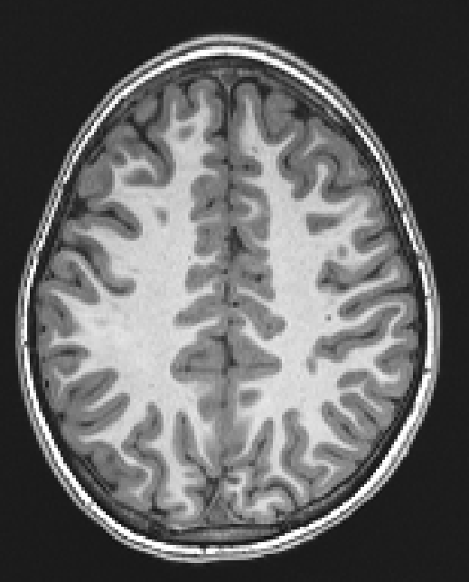
Multiple Sclerosis (MS) stands as the most prevalent chronic inflammatory disease of the central nervous system (CNS), affecting millions of people world-wide and resulting in recurring neurological symptoms impairing the proper functioning of cognitive, emotional, motor, sensory, and/or visual areas. MS occurs as a result of a person’s immune system attacking their nervous tissue, resulting in focal inflammatory lesions in the white matter (WM) of brain and spinal cord. These inflammatory lesions can be observed using magnetic resonance imaging (MRI). However, MS lesions are highly heterogeneous in terms of inflammatory responses, myelin and axonal damage, and follow different courses of microstructural and morphological evolution over time. Therefore, each lesion has a unique influence on the disability evolution of the patient. The aim of this project is to track the morphological and microstructural changes of lesions in MS patients, by characterizing their evolution using diffusion MRI. This cutting-edge imaging technique allows us to identify different microstructural compartments (such as axonal, myelin, cell infiltration, free water) in vivo, and to understand their impact on the evolution of clinical disability in MS patients. The candidate should have computational skills, have an interest in the human brain and MRI imaging, and be motivated to work in a medical context alongside medical doctors.


